Female Hip Muscles Diagram : Hip And Thigh Bones Joints Muscles Kenhub : Psoas syndrome is an injury to the iliopsoas muscle, which is a hip flexor.
byAdmin-
0
Female Hip Muscles Diagram : Hip And Thigh Bones Joints Muscles Kenhub : Psoas syndrome is an injury to the iliopsoas muscle, which is a hip flexor.. From bones to joints daniel nelson on january 1, 2019 leave a comment! The pubis, ischium, and ilium together constitute the pelvis while the thigh bone is the femur. The hip joint is made up of two bones: The sacrum and sacrotuberous ligament to the femur. Female muscle diagram and definitions.
The hip joint is made up of two bones: There are two hip bones, one on the left side of the body and the other on the right. The ball is the head of the femur (thigh bone). The adductor muscles also tend to contract when pelvic floor muscle weakness is present, as a way to compensate for the weakness. Affects athletes, particularly runners, high jumpers, and.
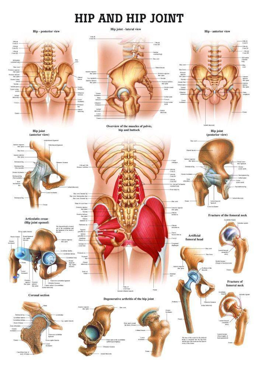
Hip And Hip Joint Laminated Anatomy Chart from cdn11.bigcommerce.com Hip anterior view, the hip is the synovial joint that connects the femur to the iliac bone. The smooth cartilage lining the socket merges into a fringe of a more. The pubis, ischium, and ilium together constitute the pelvis while the thigh bone is the femur. It is also referred to as a ball and socket joint and is surrounded by muscles, ligaments, and tendons. The ball is the head of the femur (thigh bone). It allows for complete rotations of the hip and is also. From bones to joints daniel nelson on january 1, 2019 leave a comment! Related posts of muscles of the lower back and hip diagram muscle anatomy review.
Knee feels weak despite exercise:
The groin muscles are a group of muscles situated high on the leg in the inner thigh. Browse 109 female muscle diagram stock photos and images available, or start a new search to explore more stock photos and images. The pelvis and the femur (the thighbone). There are two hip bones, one on the left side of the body and the other on the right. This muscle is located deep in the buttocks (under the gluteals). It is topographically classified as a posterior abdominal muscle but functionally as a hip muscle. It allows for complete rotations of the hip and is also. These muscles include the gluteus maximus muscle (the largest muscle in the body) and the hamstrings group, which consists of the biceps femoris, semimembranosus, and semitendinosus muscles. Both the ball and socket are lined with smooth cartilage which allows the bones to slide against each other easily (figure 1.2).; The socket in the pelvis, is called the acetabulum (figure 1.2).; Muscle anatomy review 12 photos of the muscle anatomy review anatomy muscle review quiz, cat muscle anatomy review, muscle anatomy review game, muscle review anatomy and physiology, muscular system anatomy review, human muscles, anatomy muscle review quiz, cat muscle anatomy review, muscle anatomy review game. (2017, elsevier) should be consulted. From bones to joints daniel nelson on january 1, 2019 leave a comment!
Browse 4,822 hip anatomy stock photos and images available, or search for hip replacement or knee anatomy to find more great stock photos and pictures. The four muscle of the quadriceps all extend the lower leg, and the rectus femoris additionally can flex the thigh at the hip. Related posts of muscles of the lower back and hip diagram muscle anatomy review. The hip bone is comprised of the three parts; Groin muscles help support the hip joint.
Muscles Of The Human Body Art Rocket from www.clipstudio.net The acetabulofemoral joint , commonly called the hip joint , scientifically termed is located in between the pelvis and the femur of the legs. The hip bone is comprised of the three parts; The posterior muscle group is made up of the muscles that extend (straighten) the thigh at the hip. Muscular pelvis sciatica pain and inflammation in pelvis, leg and hip. There are 3 main layers of hip abductor muscles: Affects athletes, particularly runners, high jumpers, and. They provide movement and support to the trunk. This group includes the adductor magnus, adductor longus, and adductor brevis muscles, as well as the pectineus and gracilis.
Functionally, the hip joint enjoys a very high range of motion.
Pelvic girdle and floor female pelvis and reproductive organs male pelvis and reproductive organs. Knee feels weak despite exercise: From bones to joints daniel nelson on january 1, 2019 leave a comment! Similar to learning the muscles of the lumbar spine/trunk, it can be helpful to first look at the. Human nervous system and skeleton anatomical poster. Tendinitis and bursitis many tendons around the hip connect the muscles to the joint.these tendons can easily become inflamed if you overuse them or participate in strenuous activities. It is topographically classified as a posterior abdominal muscle but functionally as a hip muscle. These muscles include the gluteus maximus muscle (the largest muscle in the body) and the hamstrings group, which consists of the biceps femoris, semimembranosus, and semitendinosus muscles. There are two hip bones, one on the left side of the body and the other on the right. (2017, elsevier) should be consulted. Human anatomy for muscle, reproductive, and skeleton. The view on the left has the rectus femoris cut away to show the vastus intermedius which is below it. The ball is the head of the femur (thigh bone).
Pelvic girdle and floor female pelvis and reproductive organs male pelvis and reproductive organs. Similar to learning the muscles of the lumbar spine/trunk, it can be helpful to first look at the. From bones to joints daniel nelson on january 1, 2019 leave a comment! The adductor muscles also tend to contract when pelvic floor muscle weakness is present, as a way to compensate for the weakness. Browse 4,822 hip anatomy stock photos and images available, or search for hip replacement or knee anatomy to find more great stock photos and pictures.
Muscles Of The Hips And Thighs Human Anatomy And Physiology Lab Bsb 141 from s3-us-west-2.amazonaws.com This group includes the adductor magnus, adductor longus, and adductor brevis muscles, as well as the pectineus and gracilis. Functionally, the hip joint enjoys a very high range of motion. The hip joint is the largest ball and socket joint in the body. Affects athletes, particularly runners, high jumpers, and. The four muscle of the quadriceps all extend the lower leg, and the rectus femoris additionally can flex the thigh at the hip. Medical flat vector illustration for clinic. Similar to learning the muscles of the lumbar spine/trunk, it can be helpful to first look at the. The sacrum and sacrotuberous ligament to the femur.
The view on the left has the rectus femoris cut away to show the vastus intermedius which is below it.
There are 3 main layers of hip abductor muscles: The hip joint is made up of two bones: Rear view of female hip and leg muscles, with labels. The groin muscles are a group of muscles situated high on the leg in the inner thigh. Groin muscles help support the hip joint. The hip itself is a ball and socket joint, much like the shoulder.the structures necessary to create this joint are the socket, the joint capsule, muscle, ligaments, and the neck. Affects athletes, particularly runners, high jumpers, and. Together, they form the part of the pelvis called the pelvic girdle. Similar to learning the muscles of the lumbar spine/trunk, it can be helpful to first look at the. In this image, you will find rectus abdominis, external oblique, inguinal ligament, tensor fascia lata, gracilis, sartorius, rectus femoris, the iliotibial band in it. Muscular pelvis sciatica pain and inflammation in pelvis, leg and hip. The quadriceps group of four muscles. The bones of the hip include the femur, the ilium, the ischium, and the pubis.